Mixed-reality application for breast-conserving surgery
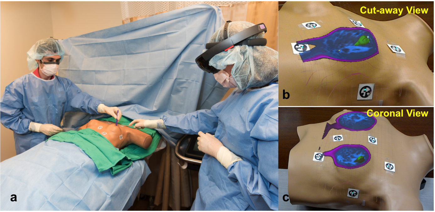
Conventional preoperative MRI and even 3D rendered MRI are presented to the surgeon on a stand-alone monitor that is separate from the patient, and requires the surgeon to mentally translate findings to the patient when planning surgery. A holoLens application was developed in collaboration with Microsoft to visualize 3D MRI overlaid onto the patient to better visualize the anatomy and potentially improve surgical planning. The holograms were perceived within a margin tolerance of < 6mm (See
ISMRM, 2017). The application was evaluated in 2 patients with palpable tumors. The tumor margins from cognitive fusion, palpation and hololens are compared (See ISMAR, 2018).
Fat-based registration of breast DCE images
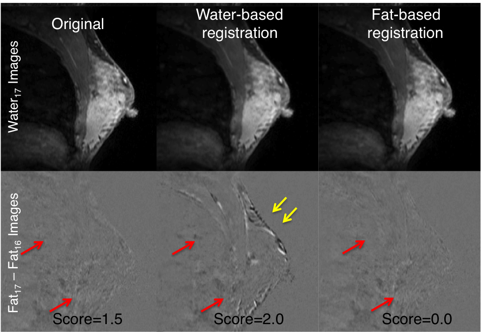
3D deformable registration of enhancing breast images is challenging even using metric such as mutual information. However the fat within the breast do not enhance and can be used to estimate deformable motion, which can be later used to deform the water images. Using a scale of 0 (no motion) to 2 (> 4 voxels of motion), the average image quality score of the fat-based registered images was 0.5±0.6, water-based registration was 0.8±0.8, and the unregistered dataset was 1.6±0.6. Fat-based registration of breast DCE images is a promising technique for performing deformable motion correction of breast without introducing new motion.(See
MRM, 2018;79(4):2408-2414)
Improved Fitting of Breast Pharmacokinetic Parameters using Dispersion models
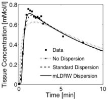
Pharmacokinetic parameters can be estimated using Tofts model by fitting the concentration-time curves (squares) acquired using dynamic contrast enhanced MRI. However, the rapid wash-in phase is not fitted well using the standard Tofts model (dotted line). The goodness-of-fit is improved by modeling delay and dispersion with the Tofts model (dashed and solid line). This also improves the sensitivity and specificity of K
trans in classifying benign and malignant tumors. (See
ISMRM, 2015)
Fast Three-Dimensional T2 weighted Prostate Imaging (3D T2-TIDE)
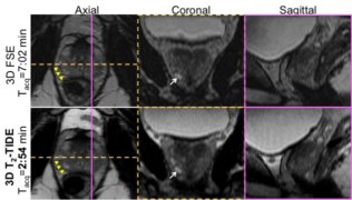
Conventional 3D fast spin echo (3D FSE) anatomic prostate imaging takes ~ 7 minutes to acquire. 3D T
2-TIDE uses variable flip angle bSSFP transient imaging for T
2 weighting and to lower the SAR at 3T. The k-space is acquired with interleaved spiral phase encode ordering for faster and efficient sampling. This reduces the acquisition duration by ~58% compared to 3D FSE with identical imaging parameters and similar image quality. (See
MRM, 2014;74(2):442-451)
Low SAR or High Contrast Variable Flip Angle Cardiac Cine bSSFP imaging
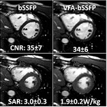
Variable Flip Angle Cardiac Cine bSSFP imaging (VFA-bSSFP) sequence using an asynchronous k-space acquisition, which is asynchronous to the cardiac cycle,
enables low SAR or high contrast cardiac cine imaging. Bloch equation simulations, phantom experiments and
in vivo imaging using VFA-bSSFP show that the SAR can be lowered by at least 36% with similar blood-myocardium contrast. VFA-bSSFP can also improve the blood-myocardium contrast by at least 28% with similar SAR compared to conventional bSSFP cardiac cine imaging. VFA-bSSFP may prove
useful for cardiac structural and functional imaging in patients with implanted devices, 3D imaging, real time imaging,
high-field imaging, and any cardiac cine application that is SAR-limited. (See
MRM, 2014;71(3):1035-1043)
Optimal Flip Angle for High Contrast Cardiac Cine bSSFP imaging
Cardiac cine imaging is routinely performed clinically for evaluation of cardiac function. High blood myocardium contrast enables clear
visualization of the chambers and segmentation of blood and myocardium for quantitative analysis of cardiac function. Bloch simulations with
perfect slice profile and stationary blood and myocardium show that maximum blood myocardium contrast can be obtained with FA of 54° at 1.5T.
However, Bloch equation simulations of flowing blood and stationary myocardium with imperfect RF slice profile show that the optimal FA for
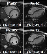
high blood myocardium contrast is 105-120°. In vivo experiments show that the optimal flip for high blood myocardium CNR,
with least artifacts is 105° for short-axis imaging plane and 75° for four chamber imaging plane. (See
MRM, 2014;73(3):1095-1103)
T2 weighted Variable flip angle PSIF imaging (T2VAPSIF)
T
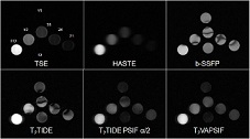
2VAPSIF sequence is based on
T2-TIDE sequence (proposed by Paul et al., ISMRM 2005). This is also
a variable flip angle acquisition in which the k-space lines up to the center are acquired using 180 flip angle and the
outer positive k-space lines are acquired with a lower flip angle. This provides fast T
2 weighted images, with lower
SAR and good edge resolution compared to the traditonal HASTE sequence. In addition, T
2VAPSIF is less sensitive to
B
0 inhomogeneities which is very useful for high and ultra high field imaging. (See
US 20130076355)
3D Black Blood Imaging
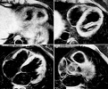
Black-blood imaging of the whole heart enables clear visualization of the cardiac chambers and the walls, cardiac masses and
other wall pathologies. We investigated the Motion Sensitized Driven Equilibrium (MSDE) preparation pulse for 3D black blood
cardiac imaging using spoiled GRE and TSE readout modules. (See
JMRI, 2012;36(2):379-386)
 Conventional preoperative MRI and even 3D rendered MRI are presented to the surgeon on a stand-alone monitor that is separate from the patient, and requires the surgeon to mentally translate findings to the patient when planning surgery. A holoLens application was developed in collaboration with Microsoft to visualize 3D MRI overlaid onto the patient to better visualize the anatomy and potentially improve surgical planning. The holograms were perceived within a margin tolerance of < 6mm (See ISMRM, 2017). The application was evaluated in 2 patients with palpable tumors. The tumor margins from cognitive fusion, palpation and hololens are compared (See ISMAR, 2018).
 3D deformable registration of enhancing breast images is challenging even using metric such as mutual information. However the fat within the breast do not enhance and can be used to estimate deformable motion, which can be later used to deform the water images. Using a scale of 0 (no motion) to 2 (> 4 voxels of motion), the average image quality score of the fat-based registered images was 0.5±0.6, water-based registration was 0.8±0.8, and the unregistered dataset was 1.6±0.6. Fat-based registration of breast DCE images is a promising technique for performing deformable motion correction of breast without introducing new motion.(See MRM, 2018;79(4):2408-2414)
 Pharmacokinetic parameters can be estimated using Tofts model by fitting the concentration-time curves (squares) acquired using dynamic contrast enhanced MRI. However, the rapid wash-in phase is not fitted well using the standard Tofts model (dotted line). The goodness-of-fit is improved by modeling delay and dispersion with the Tofts model (dashed and solid line). This also improves the sensitivity and specificity of Ktrans in classifying benign and malignant tumors. (See ISMRM, 2015)
 Conventional 3D fast spin echo (3D FSE) anatomic prostate imaging takes ~ 7 minutes to acquire. 3D T2-TIDE uses variable flip angle bSSFP transient imaging for T2 weighting and to lower the SAR at 3T. The k-space is acquired with interleaved spiral phase encode ordering for faster and efficient sampling. This reduces the acquisition duration by ~58% compared to 3D FSE with identical imaging parameters and similar image quality. (See MRM, 2014;74(2):442-451)
 Variable Flip Angle Cardiac Cine bSSFP imaging (VFA-bSSFP) sequence using an asynchronous k-space acquisition, which is asynchronous to the cardiac cycle,
enables low SAR or high contrast cardiac cine imaging. Bloch equation simulations, phantom experiments and
in vivo imaging using VFA-bSSFP show that the SAR can be lowered by at least 36% with similar blood-myocardium contrast. VFA-bSSFP can also improve the blood-myocardium contrast by at least 28% with similar SAR compared to conventional bSSFP cardiac cine imaging. VFA-bSSFP may prove
useful for cardiac structural and functional imaging in patients with implanted devices, 3D imaging, real time imaging,
high-field imaging, and any cardiac cine application that is SAR-limited. (See MRM, 2014;71(3):1035-1043)
 Cardiac cine imaging is routinely performed clinically for evaluation of cardiac function. High blood myocardium contrast enables clear
visualization of the chambers and segmentation of blood and myocardium for quantitative analysis of cardiac function. Bloch simulations with
perfect slice profile and stationary blood and myocardium show that maximum blood myocardium contrast can be obtained with FA of 54° at 1.5T.
However, Bloch equation simulations of flowing blood and stationary myocardium with imperfect RF slice profile show that the optimal FA for
high blood myocardium contrast is 105-120°. In vivo experiments show that the optimal flip for high blood myocardium CNR,
with least artifacts is 105° for short-axis imaging plane and 75° for four chamber imaging plane. (See MRM, 2014;73(3):1095-1103)
 T2VAPSIF sequence is based on T2-TIDE sequence (proposed by Paul et al., ISMRM 2005). This is also
a variable flip angle acquisition in which the k-space lines up to the center are acquired using 180 flip angle and the
outer positive k-space lines are acquired with a lower flip angle. This provides fast T2 weighted images, with lower
SAR and good edge resolution compared to the traditonal HASTE sequence. In addition, T2VAPSIF is less sensitive to
B0 inhomogeneities which is very useful for high and ultra high field imaging. (See US 20130076355)
 Black-blood imaging of the whole heart enables clear visualization of the cardiac chambers and the walls, cardiac masses and
other wall pathologies. We investigated the Motion Sensitized Driven Equilibrium (MSDE) preparation pulse for 3D black blood
cardiac imaging using spoiled GRE and TSE readout modules. (See
JMRI, 2012;36(2):379-386)